Difference between revisions of "Oral fibroma"
Jump to navigation
Jump to search
| (2 intermediate revisions by the same user not shown) | |||
| Line 1: | Line 1: | ||
{{ Infobox diagnosis | |||
| Name = {{PAGENAME}} | |||
| Image = Oral fibroma -- very low mag.jpg | |||
| Width = | |||
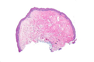
| Caption = Oral fibroma. [[H&E stain]]. | |||
| Synonyms = fibroma, focal fibrous hyperplasia, peripheral fibroma, peripheral ossifying fibroma, fibroid epulis (old term), [[fibroepithelial polyp]] | |||
| Micro = fibrous stroma +/-collagen bundles, prominent (dilated) vessels, overlying (squamous) mucosa benign +/-hyperkeratosis +/-focal ulceration | |||
| Subtypes = | |||
| LMDDx = | |||
| Stains = | |||
| IHC = | |||
| EM = | |||
| Molecular = | |||
| IF = | |||
| Gross = polypoid lesion, usually small | |||
| Grossing = | |||
| Staging = | |||
| Site = oral cavity - see ''[[oral pathology]]'' and ''[[head and neck pathology]]'' | |||
| Assdx = | |||
| Syndromes = | |||
| Clinicalhx = | |||
| Signs = | |||
| Symptoms = | |||
| Prevalence = very common | |||
| Bloodwork = | |||
| Rads = | |||
| Endoscopy = | |||
| Prognosis = benign | |||
| Other = | |||
| ClinDDx = [[squamous papilloma]], other polypoid lesions | |||
| Tx = removal | |||
}} | |||
'''Oral fibroma''', typically referred to as simply '''[[fibroma]]''', is a very common benign lesion in [[oral pathology]]. | '''Oral fibroma''', typically referred to as simply '''[[fibroma]]''', is a very common benign lesion in [[oral pathology]]. | ||
| Line 21: | Line 53: | ||
*Overlying (squamous) mucosa benign (flat). | *Overlying (squamous) mucosa benign (flat). | ||
**+/-Hyperkeratosis +/-focal ulceration.<ref name=Ref_HaNP240>{{Ref HaNP|240}}</ref> | **+/-Hyperkeratosis +/-focal ulceration.<ref name=Ref_HaNP240>{{Ref HaNP|240}}</ref> | ||
===Images=== | |||
<gallery> | |||
Image: Oral fibroma -- very low mag.jpg | OF - very low mag. (WC) | |||
Image: Oral fibroma -- low mag.jpg | OF - low mag. (WC) | |||
Image: Oral fibroma -- intermed mag.jpg | OF - intermed. mag. (WC) | |||
</gallery> | |||
==Sign out== | ==Sign out== | ||
Latest revision as of 19:10, 3 February 2016
| Oral fibroma | |
|---|---|
| Diagnosis in short | |
 Oral fibroma. H&E stain. | |
|
| |
| Synonyms | fibroma, focal fibrous hyperplasia, peripheral fibroma, peripheral ossifying fibroma, fibroid epulis (old term), fibroepithelial polyp |
|
| |
| LM | fibrous stroma +/-collagen bundles, prominent (dilated) vessels, overlying (squamous) mucosa benign +/-hyperkeratosis +/-focal ulceration |
| Gross | polypoid lesion, usually small |
| Site | oral cavity - see oral pathology and head and neck pathology |
|
| |
| Prevalence | very common |
| Prognosis | benign |
| Clin. DDx | squamous papilloma, other polypoid lesions |
| Treatment | removal |
Oral fibroma, typically referred to as simply fibroma, is a very common benign lesion in oral pathology.
It is also known as focal fibrous hyperplasia, peripheral fibroma, peripheral ossifying fibroma, fibroid epulis (old term), and fibroepithelial polyp.[1][2][3]
General
- Most common oral cavity tumour.[3]
- Female predominance (female:male = 2:1), usually 30-50 years old.[3]
- Multiple oral fibromas may be seen in Cowden disease.[4][5]
- Histologically similar to fibrous papule.[6]
Clinical DDx:
Microscopic
Features:[6]
- Fibrous stroma - key feature.
- "Very pink" at low power.
- +/-Collagen bundles, may be prominent.
- Prominent (dilated) vessels.
- Overlying (squamous) mucosa benign (flat).
- +/-Hyperkeratosis +/-focal ulceration.[3]
Images
Sign out
Lower Buccal Lesion, Left, Excision:
- Fibroma.
- NEGATIVE for malignancy.
Left Tongue Lesion, Excision:
- Fibroma with overlying squamous epithelium with parakeratosis.
- NEGATIVE for malignancy.
Block letters
TONGUE LESION, BIOPSY:
- FIBROMA.
Micro
The sections show a polypoid fragment of squamous mucosa with a thin layer of parakeratosis. The subepithelial tissue consists of abundant fibrous tissue and has dilated small blood vessels.
No significant inflammation is present. No nuclear atypia is apparent. No necrosis is present. No proliferative activity is apparent.
See also
References
- ↑ Mills, Stacey E; Carter, Darryl; Greenson, Joel K; Reuter, Victor E; Stoler, Mark H (2009). Sternberg's Diagnostic Surgical Pathology (5th ed.). Lippincott Williams & Wilkins. pp. 775. ISBN 978-0781779425.
- ↑ URL: http://emedicine.medscape.com/article/1080948-overview#aw2aab6b3. Accessed on: 20 August 2012.
- ↑ 3.0 3.1 3.2 3.3 Thompson, Lester D. R. (2006). Head and Neck Pathology: A Volume in Foundations in Diagnostic Pathology Series (1st ed.). Churchill Livingstone. pp. 240. ISBN 978-0443069604.
- ↑ Segura Saint-Gerons, R.; Ceballos Salobreña, A.; Toro Rojas, M.; Gándara Rey, JM. (Aug 2006). "Oral manifestations of Cowden's disease. Presentation of a clinical case.". Med Oral Patol Oral Cir Bucal 11 (5): E421-4. PMID 16878060.
- ↑ Oliveira, MA.; Medina, JB.; Xavier, FC.; Magalhães, M.; Ortega, KL. (2010). "Cowden syndrome.". Dermatol Online J 16 (1): 7. PMID 20137749.
- ↑ 6.0 6.1 Fernandez-Flores, A. (Jul 2010). "Solitary oral fibromas of the tongue show similar morphologic features to fibrous papule of the face: a study of 31 cases.". Am J Dermatopathol 32 (5): 442-7. doi:10.1097/DAD.0b013e3181c47142. PMID 20421776.


