Difference between revisions of "Pneumothorax"
Jump to navigation
Jump to search
| Line 3: | Line 3: | ||
==General== | ==General== | ||
*[[Clinical diagnosis]] | *[[Clinical diagnosis]] by radiology ''or'' [[autopsy]] diagnosis. | ||
===Causes=== | ===Causes=== | ||
Revision as of 13:36, 2 February 2017
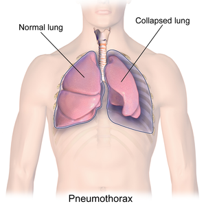
Pneumothorax is air within the potential space between the parietal pleura and visceral pleura leading to a partial or complete collapse of the lung.
General
- Clinical diagnosis by radiology or autopsy diagnosis.
Causes
- Primary.[1]
- No underlying cause.
- Secondary - underlying disease.
- Emphysema.
- Bullous disease.
- Blunt force trauma - esp. rib fractures.
- Interstitial lung disease.[citation needed]
- Iatrogenic.
- Lymphangioleiomyomatosis.
Sign out
A. Lung, Right Upper Lobe - Apical Segment, Wedge Resection:
- Mild emphysematous changes.
- Focal subpleural fibrosis.
- NEGATIVE for significant inflammation.
- NEGATIVE for significant interstitial fibrosis.
- NEGATIVE for malignancy.
- Please see comment.

B. Lung, Right Upper Lobe - Posterior Segment, Wedge Resection:
- Mild emphysematous changes.
- Focal subpleural fibrosis.
- NEGATIVE for significant inflammation.
- NEGATIVE for significant interstitial fibrosis.
- NEGATIVE for malignancy.
- Please see comment.

Comment:
The wedge-shaped pattern of fibrotic healing seen in the pleura is typical
of spontaneous pneumothorax.

The emphysema present may be a contributory factor; however, other causes
must be excluded clinically.
See also
References
- ↑ Papagiannis, A.; Lazaridis, G.; Zarogoulidis, K.; Papaiwannou, A.; Karavergou, A.; Lampaki, S.; Baka, S.; Mpoukovinas, I. et al. (Mar 2015). "Pneumothorax: an up to date "introduction".". Ann Transl Med 3 (4): 53. doi:10.3978/j.issn.2305-5839.2015.03.23. PMID 25861608.
