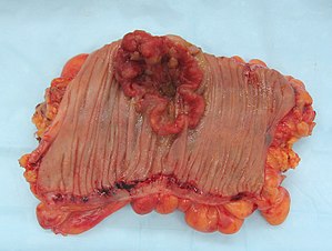
Cancer
Jump to navigation
Jump to search
Cancer is a leading cause of mortality and morbidity. It is what keeps pathologists busy.
This article deals with general concepts in cancer. Specific forms of cancer are dealt with in their own articles (e.g. colorectal adenocarcinoma) and/or the anatomical site articles.
In addition to be an introduction to cancer, this article also deals with cancers of unknown primary.
Definition of cancer
The Robbins definition of cancer:[1]
- Tumour with the capacity to:
- Invade (infiltrate) and destroy adjacent tissue.
- Spread to other sites (metastasize).
Note:
- Tumour = abnormal mass of tissue.[1]
- Neoplasm = literally "new growth"; used interchangeably with tumour.[1]
Tumours
Tumours per Robbins[1] consist of:
- Clonal expansions.
- Stromal components - controlled by the clonal component.
Note:
- Tumour may be used to refer to a non-neoplastic process; one dictionary definition of tumour is abnormal swelling.[2] Also see note in definition of cancer.
Clinical features of cancer
- Uncontrolled growth (compared to normal tissue).
- Autonomous growth - without external stimulus.
- Direct invasion/destruction of tissue.
- Metastases.
Notes:
- 3 may or many not be present. Primary brain cancer rarely metastasizes.
Theory
- Cancer results from an accumulation of driver mutations.
- It is thought that approximately seven driver mutations are required.[3]
Cancers of unknown primary
- Undifferentiated malignancy, unknown primary tumours, and poorly differentiated malignancy redirect here.
Clinical history
- Past history of cancer, i.e. is it a recurrence?
- Family history of cancer?
- Symptoms/presentation.
- Occupation & documented exposures - esp. smoking, excessive alcohol use.
- Impression from clinician(s).
Radiology/gross pathology
- Where is the largest lesion?
- Location of the other lesions?
- Is the pattern of spread compatible with the suspected primary site?
- Is the tumour at a location of a common (primary) cancer in the individual's demographic group?
Pathologic features
- Histomorphologic characteristics - see modified general morphologic DDx of malignancy.
- Special stains.
- Immunohistochemistry.
- Molecular pathology.
- Electron microscopy.
Incidence and deaths in Canada
Men
Incidence:[4]
- Prostate.
- Lung.
- Colorectal.
- Bladder.
- Non-Hodgkin lymphoma.
- Kidney.
- Melanoma.
- Leukemia.
- Oral.
- Pancreas.
- Stomach.
- Brain.
Death:[4]
- Lung.
- Colorectal.
- Prostate.
- Pancreas.
- Non-Hodgkin lymphoma.
- Leukemia.
- Esophagus.
- Bladder.
- Stomach.
- Kidney.
- Brain.
Women
Incidence:[4]
- Breast.
- Lung.
- Colorectal.
- Body of uterus.
- Thyroid.
- Non-Hodgkin lymphoma.
- Ovary.
- Melanoma.
- Pancreas.
- Leukemia.
- Kidney.
- Bladder.
- Cervix.
Death:[4]
- Lung.
- Breast.
- Colorectal.
- Pancreas.
- Ovary.
- Non-Hodgkin lymphoma.
- Leukemia.
- Body of uterus.
- Brain.
- Stomach.
- Multiple myeloma.
- Kidney.
- Bladder.
See also
References
- ↑ 1.0 1.1 1.2 1.3 Mitchell, Richard; Kumar, Vinay; Fausto, Nelson; Abbas, Abul K.; Aster, Jon (2011). Pocket Companion to Robbins & Cotran Pathologic Basis of Disease (8th ed.). Elsevier Saunders. pp. 145. ISBN 978-1416054542.
- ↑ URL: http://dictionary.reference.com/browse/tumour?s=t. Accessed on: 22 May 2012.
- ↑ Nordling C (1953). "A new theory on cancer-inducing mechanism". Br J Cancer 7 (1): 68–72. doi:10.1038/bjc.1953.8. PMC 2007872. PMID 13051507. http://www.carlonordling.se/Cancer.html.
- ↑ 4.0 4.1 4.2 4.3 URL: http://www.cancer.ca/Canada-wide/About%20cancer/Cancer%20statistics/~/media/CCS/Canada%20wide/Files%20List/English%20files%20heading/Powerpoint%20-%20Policy%20-%20Canadian%20Cancer%20Statistics%20-%20English%202011/2011%20Figures%201.1-3.2%20E.ashx. Accessed on: 24 September 2011.
